C4.5: Electron Microscopy Studies of the Properties of Nanoparticles
Subproject Leader: Dagmar Gerthsen
Laboratorium für Elektronenmikroskopie (LEM), KIT
Contributing Scientists: Radian Popescu, Eric Prestat, Reinhard Schneider
Electron microscopic techniques are applied in subproject C4.5 to study the structural and electronic properties of metallic clusters and novel nanoparticles. Furthermore, time-dependent investigations are carried out to monitor cluster dynamics and analyse the (in)stability of cluster-size distributions of mass and non-mass-selected clusters which is of significant interest for numerous applications.
Coarsening of the Size Distribution of Metallic Clusters
Using transmission electron microscopy (TEM) and transmission electron holography, the redistribution non-size-selected Au-clusters was studied [1]. The samples were produced by laser deposition on amorphous carbon (a-C) substrates in subproject C4.6. After 3-4 months storage under ambient conditions, i.e. at room temperature in air, the formation of comparably large Au-islands with embedded Au-clusters is observed. A model potential for the interaction of Au-atoms with an a-C substrate was obtained by density functional calculations. The potential was used in subsequent Monte-Carlo simulations which describe the island formation process and the shape of the embedded Au-clusters.
In another study, coarsening of mass-selected Aun (n=4, 6, 13 and 20) clusters on amorphous carbon was investigated [2]. The samples were stored under ambient conditions for time periods of up to 32 months. A significant increase of the cluster sizes is observed which can be described by surface Ostwald ripening (OR) while Smoluchowski ripening, i.e. coalescence of clusters, is suppressed by strong bonding of the clusters to the amorphous carbon substrate. A detailed analysis of surface diffusion coefficient suggests that the rate of surface OR for mass-selected Aun-clusters increases with the cluster size in the sequence Au4°≤°Au6°<°Au13°<°Au20. The activation of the coarsening process is rationalized by initial variations of the cluster sizes due to the deposition process itself and/or the interaction of the clusters with the substrate. Moreover, the presence of initial deposited Au-clusters as different isomers with slightly different chemical potential on the substrate, may also initiate the coarsening. Furthermore, we find that the coarsening is most pronounced for the paucidispersed sample with Aum (10 ≤ m ≤ 20) clusters. A possible explanation of this behaviour is the presence of an initial distribution of different cluster sizes directly after deposition.
Mean Inner Coulomb Potential of Au-Clusters
The mean inner Coulomb potential (MIP) is a basic material property, which, e.g., determines the amplitude of the electron wave scattered in forward direction in TEM and other electron scattering experiments. The mean inner potential can be determined by electron holography in a transmission electron microscope which allows reconstruction of the amplitude and phase of the electron wave. A strong increase of the MIP of Au-clusters is observed with decreasing cluster size. This effect can be explained by the large surface-to-volume ratio of clusters, the reduction of bond length of surface atoms and the corresponding increase of electron density in the cluster. The increase of the MIP leads to a significantly increased amplitude of electron wave scattered in forward direction which needs to be taken into account for quantification of TEM data [3].
Structure of Novel Nanoparticles
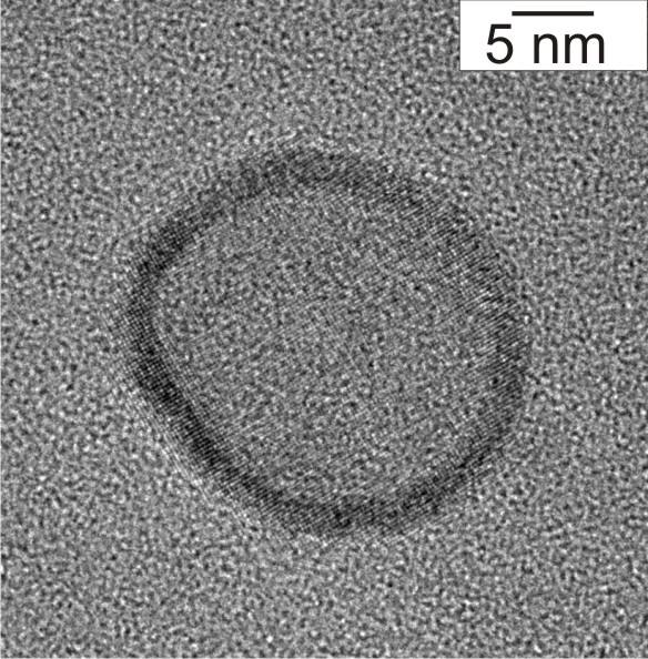
In subproject C4.5 we also analyse the structure and chemical properties of novel nanoparticles which are synthesized by other CFN groups. One focus of project C1.4 is the synthesis of metall and metal-oxide hollow nanoparticles. Fig. 1 shows as an example a high-resolution transmission electron microscopy (HRTEM) image of a hollow gold sphere with a wall thickness of only 2 - 3 nm [4].
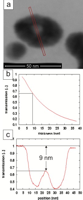
The reconstruction of the three-dimensional shape of hollow irregularly shaped Ag-particles (Fig. 2a) was accomplished by quantitative low-energy scanning transmission electron microscopy (low-energy STEM) in the bright-field mode [5]. In this work, the local wall thickness in electron-beam direction was determined along the line in Fig. 2a by comparison of the experimental STEM intensity (Fig. 2c) with calculated STEM intensities using Monte-Carlo simulations (Fig. 2b).
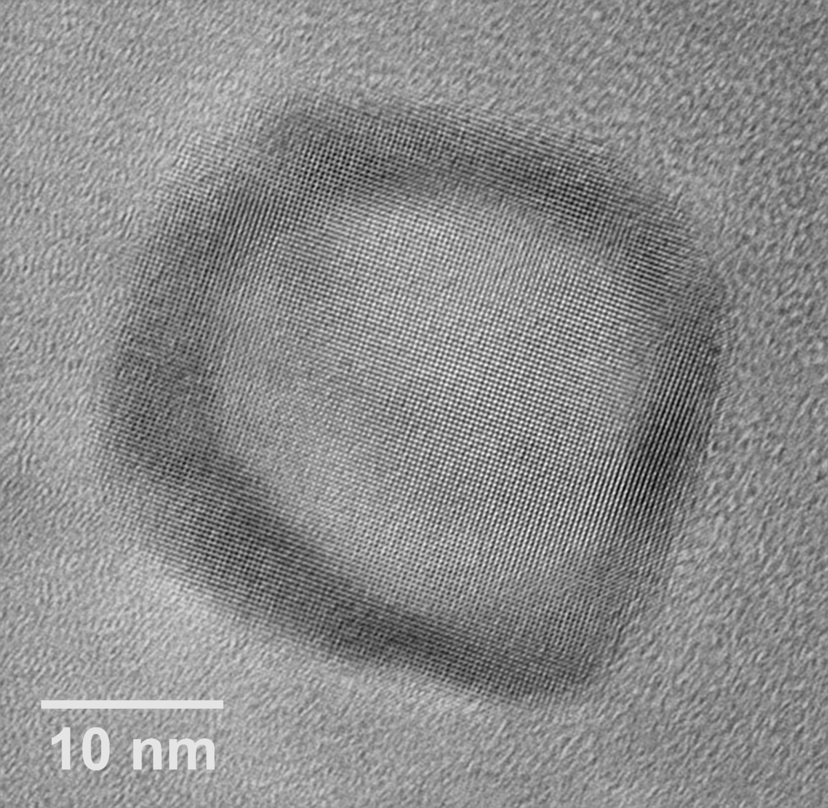
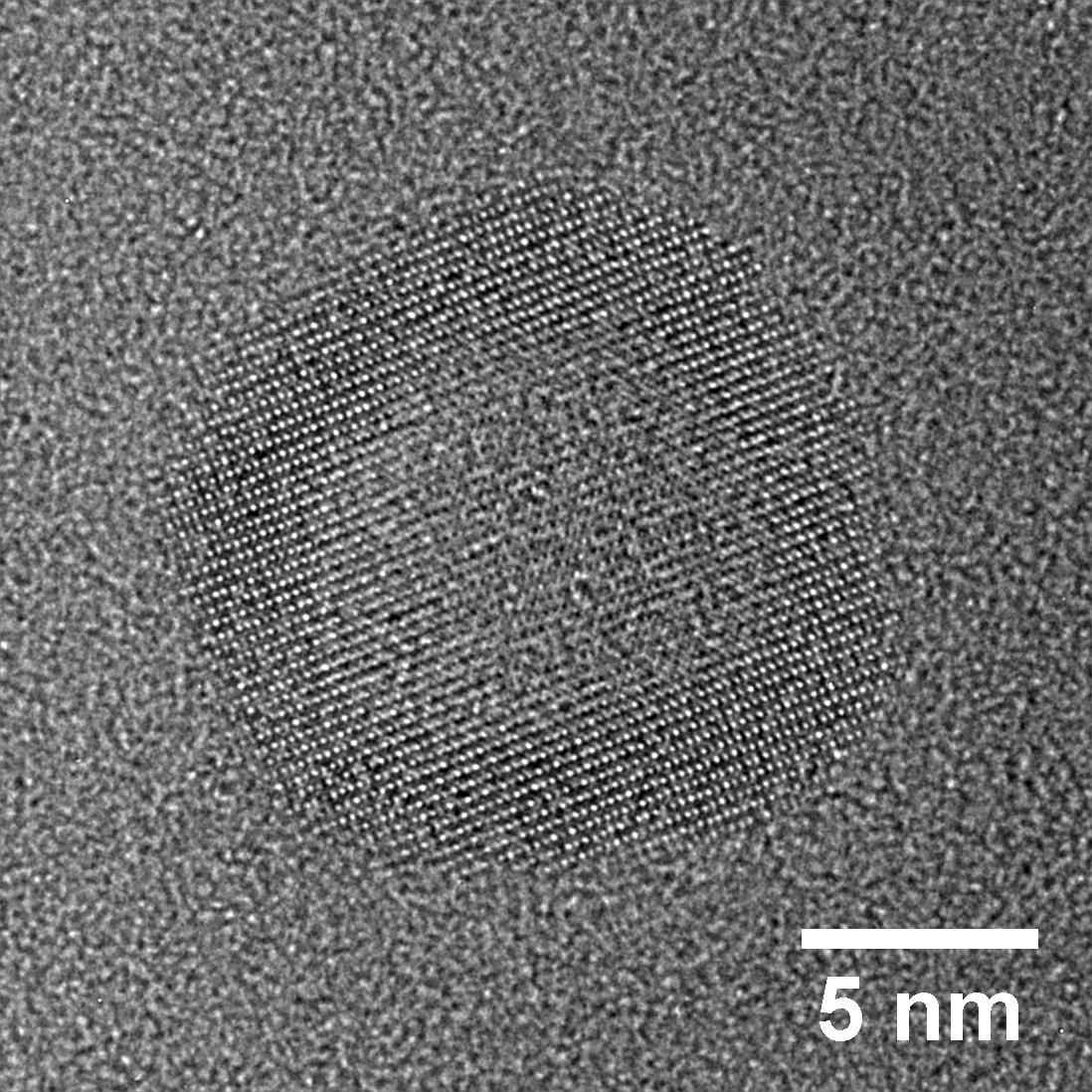
Numerous novel hollow oxide nanoparticles synthesized in project C1.4 were characterized by HRTEM consisting of e.g. ZnO (Fig. 3) or Fe3O4 (Fig. 4).
References
|
[1] |
M. Wanner, R. Werner, and D. Gerthsen, Dynamics of gold clusters on amorphous carbon films induced by annealing in a transmission electron microscope, Surf. Sci. 600, 632 (2006) |
|
[2] |
R.Popescu, R. Schneider, D. Gerthsen, A. Boettcher, D. Loeffler, P. Weis, M. M. Kappes, Coarsening of mass-selected Au clusters on amorphous carbon at room temperature, Surf. Sci. 603, 3119 (2009) |
|
[3] |
R. Popescu, E. Müller, D.Gerthsen, M. Wanner, M. Schowalter, A. Rosenauer, A. Böttcher, D. Löffler, P. Weis, Increase of the mean inner Coulomb potential in Au clusters induced by surface tension and its implication for electron scattering, Phys. Rev. B 76, 235411 (2007) |
|
[4] |
C. Zimmermann, C. Feldmann, M. Wanner and D. Gerthsen, Nanoscale Gold Hollow Spheres Through a Microemulsion Approach, Small 3, 1347 (2007) |
|
[5] |
C. Kind, R. Popescu, E. Müller, D. Gerthsen, and C. Feldmann, Microemulsion-based synthesis of nanoscaled silver hollow spheres and direct comparison with massive particles of similar size, Nanoscale 2, 2223 (2010) |
List of Publications 2006-2011 as PDF
Subproject Report 2006-2010 as PDF