C3.2: Frequency and Time-Domain Electronic Spectroscopy of Isolated Clusters and Carbon Nanotubes
Subproject Leader:
Manfred Kappes
Institut für Physikalische Chemie, KIT
Andreas Unterreiner
Institut für Physikalische Chemie, KIT
Contributing Scientists:
Present: Melanie Klinger, Yu Liang, Seyithan Ulas, Matthias Vonderach
Past: Thomas Wolf, Carolin Blum, Rebecca Kelting

Subproject C3.02: "Frequency and Time-Domain Electronic Spectroscopy of Isolated Clusters and Carbon Nanotubes" concentrates on the characteristic interactions of such molecular nanostructures with light, on the modulation of such interactions via external perturbations - ideally controllable at the single molecule level – as well as on electronic decay processes following photoexcitation – down to the time scale of tens of femtoseconds. We study the same molecular nanostructures, for instance, single-walled carbon nanotubes and fullerenes, by different spectroscopic tools, both in the frequency and time domain. Furthermore, different environments (external pertubations) are realized for the the same chromophores, ranging from fully isolated in gas-phase (under ultrahigh vacuum) to surface bound or enclosed in condensed phase (e.g., dispersed in liquids).
Frequency-Domain Spectroscopy of Single-Walled Carbon Nantotubes (SWNTs)
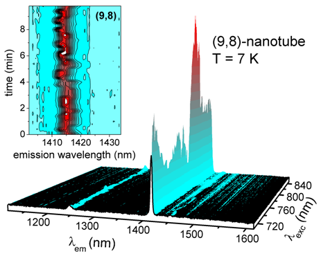
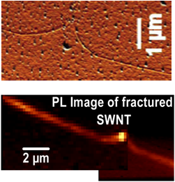
A number of frequency-domain spectroscopic techniques has been applied/ developed within the subproject, primarily for investigations of SWNTs down to individual species. They include a near-infrared (NIR) photoluminescence (PL) laser microscope with a scanning excitation wavelength of ~600-1000 nm and a Raman microscope with detection of Raman signals down to ~10/20 cm-1 Stokes/ anti-Stokes shift. These instruments provide a spatial/lateral resolution down to ~300 nm. A dedicated microscope for very fast anisotropic (polarisation-modulated) Rayleigh imaging of individual nanotubes is under development. Using these spectroscopic tools, various SWNT samples have been characterized and different nanotube properties have been investigated. Two examples are presented below. Fig.1 compares characteristic PL “blinking” (temporal fluctuations in intensity and spectral position) of individual SWNTs in an (organic) matrix with stable emission from suspended (matrix-free) nanotubes. PL “blinking” has been observed for various types of molecular and nanostructured fluorophores, in particular at low temperatures; its exact mechanism(s) is still a matter of research. In the case of SWNTs, PL “blinking” appears not to be an intrinsic property, instead it is caused by interactions with the nanotube environment [1]. Fig. 2 illustrates the complimentary use of atomic force microscopy (AFM) and PL imaging to characterise SWNTs grown as well-aligned mm-long arrays on Si/SiO2 substrates (cooperation with Prof. Yan Li, Peking University). An AFM tip can also be used for controllable manipulation (bending, cutting) of nanotubes on surfaces. This typically results in residual mechanical stresses in the nanotube, e.g., in the vicinity of the fracture site. The stress can be readily detected with a PL microscope via characteristic shifts of the electronic transition energies and, consequently, of the PL peaks [2].
Chemical and Physical Fractionation of Single-Walled Carbon Nanotubes
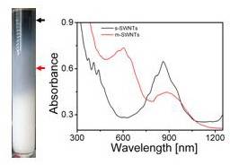
Typical as-prepared SWNT materials are quite inhomogeneous, containing a variety of SWNT species (defined by chiral indices (n, m)), their aggregates (bundles) as well as impurities. Well-defined samples are important not only for spectroscopic studies but, probably even more so, for various potential applications of SWNTs. Therefore, this subproject also includes the investigation and development of efficient separation methods for SWNTs. To date, several monochiral ((n, m)-pure) or highly purified SWNT samples have become available by polymer wrapping. Furthermore we have developed a procedure which allows for large scale fractionation of metallic vs. semiconducting SWNT samples (Fig. 3) [3].
Our ultrafast laser spectroscopic investigations focus on electronic relaxation processes in molecular nanostructures including nanotubes in solution. Such systems are excited and probed in the spectral range from UV to NIR, with sub-100 fs time resolution. For instance, time-resolved one- and two-color pump-probe experiments on (9,7)-selected SWNTs dispersed in toluene have yielded new insights into relaxation pathways of the first excited electronic state, E11. In fact, excitation into this region accesses a staircase of “bright” and “dark” excitonic states, with a complicated pattern of relaxation pathways – significantly defining the optical properties of nanotubes, e.g., their photoluminescence efficiency. In our experiments NIR probes allowed the direct observation of a “dark” state in the vicinity of the first “bright” exciton. Population of this dark state occurs within 1 ps after optical excitation. Further investigations have comprised, for instance, a measurement of the ultrafast relaxation dynamics of fullerene anions in solution by the technique of transient anisotropy. Among other interesting properties, fullerene anions can demonstrate unusual and extremely fast changes of their transition dipole moments, which have been ascribed to pseudo-rotations of fullerene cages distorted by the Jahn-Teller effect [4].
Time-Resolved Photoelectron Spectroscopy (TRPES) of Isolated Anions
TRPES is a powerful method to access electronic and geometrical structure, stability as well as electronic and vibrational relaxation for charged molecular species isolated in the gas phase, i.e. under “surrounding-free” conditions. Note that the same species (chromophores) may also be studied in condensed phase, thus elucidating effects of the surroundings. Within this subproject, a TRPE spectrometer has been developed/built and coupled to the fs-pump-probe multi-color laser facility. In this spectrometer, molecular anions are electrosprayed from solution, mass selected, transferred, and decelerated to reduce Doppler broadening - prior to photoexcitation and subsequent photoelectron detachment. A magnetic bottle spectrometer has been implemented for efficient collection of photoelectrons. For instance, TRPE spectroscopy has been applied to photoinduced unimolecular fragmentation of hexabromo-iridiate (IV) dianions, IrBr62- [5] and to the photoinduced excited state tunneling autodetachment of metal phthalocyanine tetrasulfonate tetraanions [6].
References
| [1] | O. Kiowski, S. Lebedkin, F. Hennrich, and M. Kappes, Phys. Rev. B, 76, 075422 (2007) |
| [2] | O. Kiowski, S.-S. Jester, S. Lebedkin, Z. Jin, Y. Li and M. Kappes, Phys. Rev. B, 80, 075426 (2009) |
| [3] | F. Hennrich, K. Moshammer, and M. Kappes, Nano Research, 2, 593 (2009) |
| [4] | O. Schalk and A.-N. Unterreiner, Phys. Chem. Chem. Phys., 12, 655 (2010) |
| [5] | C. Rensing, O.T. Ehrler, J.-P. Yang, A.-N. Unterreiner and M. M. Kappes, J. Chem. Phys., 130, 234306 (2009) |
List of Publications 2006-2011 as PDF
Subproject Report 2006-2010 as PDF